

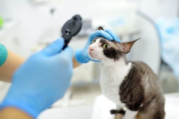
Observing a sudden shift in how your dog or cat navigates their home environment can be a deeply alarming experience for any companion owner. One day your pet moves around living room furniture with absolute confidence, and the next, they hesitate at doorway thresholds, misjudge sofa edges, or bump into doorframes. When optical changes develop rapidly, many owners immediately search for reliable vets near me to understand what is happening behind their pet’s eyes.
One of the most serious underlying conditions responsible for rapid visual decline in companion animals is feline and canine glaucoma. This condition involves a sudden build-up of fluid pressure within the eye socket, which rapidly damages delicate optic nerve fibers and retinal tissue. Seeking urgent pet care ensures that clinical teams can evaluate intraocular pressure immediately, relieving pain and preserving remaining sight. For families living in Brooklyn, recognizing the early warning signs of elevated eye pressure helps ensure companion animals receive swift, specialized medical attention.
The Biological Mechanisms of Glaucoma and Vision Loss
To understand how glaucoma impacts an animal’s eyesight, it helps to explore how fluid normally moves within the healthy eye structure.
- Fluid Production and Drainage Balance:The eye constantly produces a clear fluid called aqueous humor to nourish internal lenses and maintain shape. In a healthy eye, this fluid drains out through a complex microscopic meshwork where the iris meets the cornea.
- Drainage Blockage and Pressure Spikes:When inflammatory debris, lens dislocations, or inherited structural narrowness block these drainage pathways, fluid accumulates continuously. Intraocular pressure spikes dramatically within hours, placing immense mechanical stress on the back of the eye.
- Optic Nerve and Retinal Damage:High intraocular pressure crushes delicate nerve fibers connecting the eye to the brain while restricting blood flow to the retina. Without rapid pressure-reducing treatment, permanent loss of functional vision can occur within a brief window of time.
Key Warning Signs: Home Observation vs. Clinical Symptoms
Distinguishing between minor age-related eye haziness and acute glaucoma requires watching for physical discomfort alongside navigational changes.
Signs Safe for Temporary At-Home Observation
If your pet exhibits any of these mild, non-painful variations while maintaining bright energy and confident movement, brief home monitoring is appropriate:
- Mild, symmetrical bluish lens haziness in an aging senior pet without any squinting
- Minor, clear watery eye tearing after playing outdoors on a windy afternoon
- Intact navigational confidence when walking through both brightly lit and dark spaces
- Smooth, shiny corneal surfaces with zero visible swelling, redness, or lid spasms
Severe Signs Demanding Immediate Urgent Vet Care
If your companion displays signs of acute ocular pressure or sudden visual impairment, seeking intermediate medical evaluation prevents permanent tissue damage:
- Sudden cloudiness, a diffuse bluish-gray film, or a steamed-glass appearance across the cornea
- Widely dilated pupils that fail to constrict when exposed to bright indoor lights
- Prominent, engorged blood vessels visible across the white sclera and surrounding tissues
- Persistent squinting, keeping an eye held tightly shut, or frequent face rubbing
- Bumping into furniture, hesitation before staircases, or obvious spatial confusion
Point-of-Care Diagnostics for High Ocular Pressure
When you bring your pet in for clinical evaluation regarding suspected eye pressure, veterinary teams utilize non-invasive point-of-care tools to inspect eye structures without delay.
- Ophthalmic Tonometry Measurement: Uses a gentle, non-invasive electronic probe to measure intraocular fluid pressure in millimeters of mercury within seconds.
- Gonioscopic Drainage Angle Exam: Inspects the microscopic drainage channels in the anterior chamber using specialized magnifying lenses.
- Fluorescein Dye Application: Rules out underlying corneal surface scratches or ulcers before initiating pressure-reducing therapies.
Combining instant diagnostic feedback with pet-safe topical eye drops, oral pressure-reducing medications, and hyperosmotic therapies halts pain at its source and works to preserve remaining sight.
Supporting Companion Animals Across Brooklyn
Protecting your pet’s visual health requires attentive daily care, routine checks, and staying mindful of subtle changes in their behavior. Checking your companion’s eyes during daily petting sessions allows you to spot minor cloudiness, pupil asymmetry, or surface redness before mobility is affected.
If you are from Brooklyn and looking for urgent vet care, taking fast action at the first sign of eye cloudiness, redness, or sudden disorientation provides your pet with the best opportunity for a smooth, full recovery.
Providing timely medical attention when health changes occur preserves your companion’s quality of life and offers complete peace of mind for your entire household.
Taking Action for Your Pet’s Visual Comfort
When your dog or cat exhibits sudden navigational changes, eye cloudiness, or physical discomfort, securing a prompt evaluation keeps their recovery moving in the right direction. You can schedule an online appointment through our portal to reserve a convenient time for your pet to be examined by our medical staff.
If your companion is actively displaying uncomfortable eye symptoms or sudden optical changes and you prefer to speak directly with our clinical staff before arriving, please call our team at 718-387-0541 so we can offer immediate guidance and prepare for your visit.
Our dedicated medical team looks forward to delivering gentle, expert care for your companion at Northside Veterinary Clinic.
Frequently Asked Questions
Q1. Is glaucoma in dogs and cats a painful condition?
Ans: Yes. Elevated intraocular pressure causes a continuous, intense headache and localized eye pain. Pets experiencing high eye pressure often become unusually quiet, hide in dark areas, refuse food, show sensitivity to light, or squint persistently as they try to manage the continuous discomfort.
Q2. Can glaucoma affect both eyes at the same time?
Ans: Primary inherited glaucoma often develops in one eye first, but the opposite eye remains at very high risk for similar pressure spikes over time. Secondary glaucoma caused by internal eye inflammation, cataracts, or trauma typically affects only the injured or inflamed eye initially.
Q3. How quickly can high intraocular pressure cause permanent vision loss?
Ans: Severe intraocular pressure spikes can cause permanent, irreversible damage to the optic nerve and retinal cells in as little as 24 to 48 hours. Because the timeline for saving functional sight is brief, immediate veterinary evaluation and pressure testing are strictly necessary.
Q4. Are certain breeds of dogs and cats more prone to developing glaucoma?
Ans: Yes. Certain canine breeds including Cocker Spaniels, Basset Hounds, Boston Terriers, Chow Chows, and Beagles possess an anatomical predisposition for primary glaucoma. Feline breeds such as Siamese and Burmese cats can also experience genetic or secondary pressure elevations.
Q5. How should I safely transport a pet experiencing eye pain to the clinic?
Ans: Keep your vehicle cool, quiet, and shaded from bright sunlight to minimize light sensitivity. Place cats and small dogs inside a soft, padded carrier lined with familiar blankets. For larger dogs, use a short leash with calm vocal encouragement, avoiding any pressure on the neck or head.
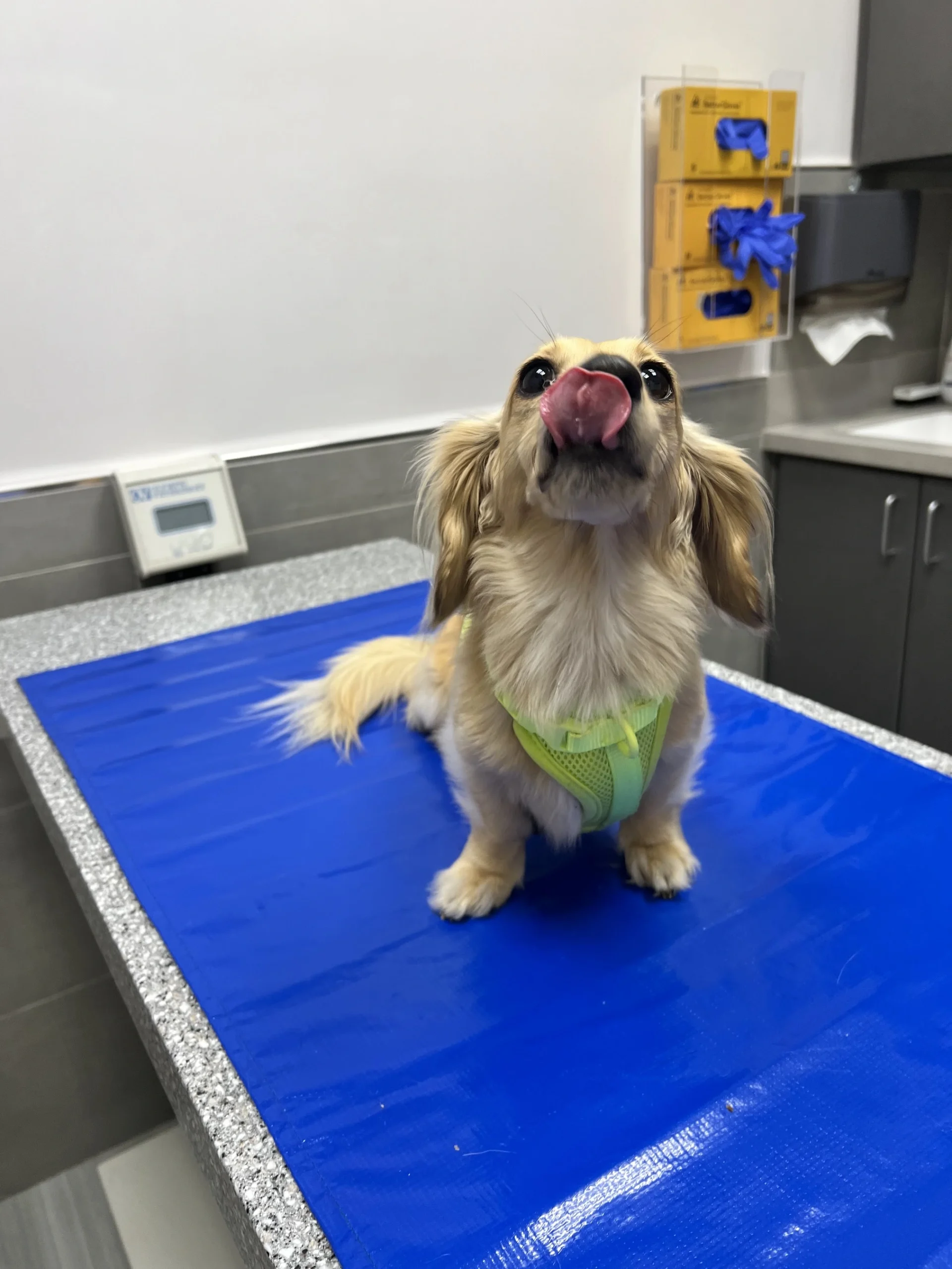
Find a Puppy & Kitten Vet Near Brooklyn, New York
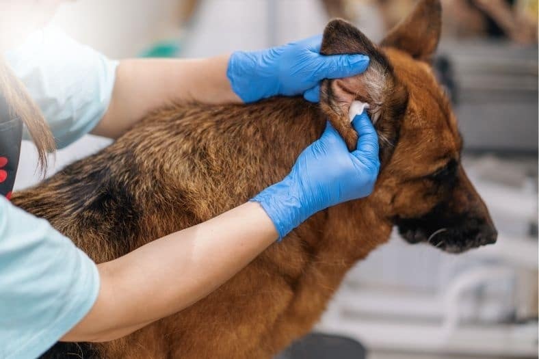
Finding a full service vet clinic in Brooklyn for your new furry friend is easy with our online direct booking feature. Both new and existing clients seeking care from a veterinarian may schedule an appointment at Northside Veterinary Clinic.